
fMRI Studies of Writing Processes in the Brain
fMRI studies reveal how our brains function during writing tasks. Here's what you need to know:
- Writing engages multiple brain regions, primarily in the left hemisphere
- Key areas include the premotor cortex, parietal cortex, and fusiform gyrus
- Handwriting activates more brain regions than typing
- Good writers show more efficient brain activation patterns
- fMRI helps diagnose writing disorders like dysgraphia and dyslexia
Recent advances in fMRI technology allow for faster, higher-resolution brain imaging, opening new avenues for writing research.
| Brain Region | Function in Writing |
|---|---|
| Left premotor cortex | Motor control |
| Left superior parietal cortex | Spatial processing |
| Left fusiform gyrus | Visual word recognition |
| Exner's area | Writing movement memory |
These insights are reshaping our understanding of writing processes and could lead to better educational strategies and treatments for writing disorders.
Related video from YouTube
How fMRI works
fMRI basics
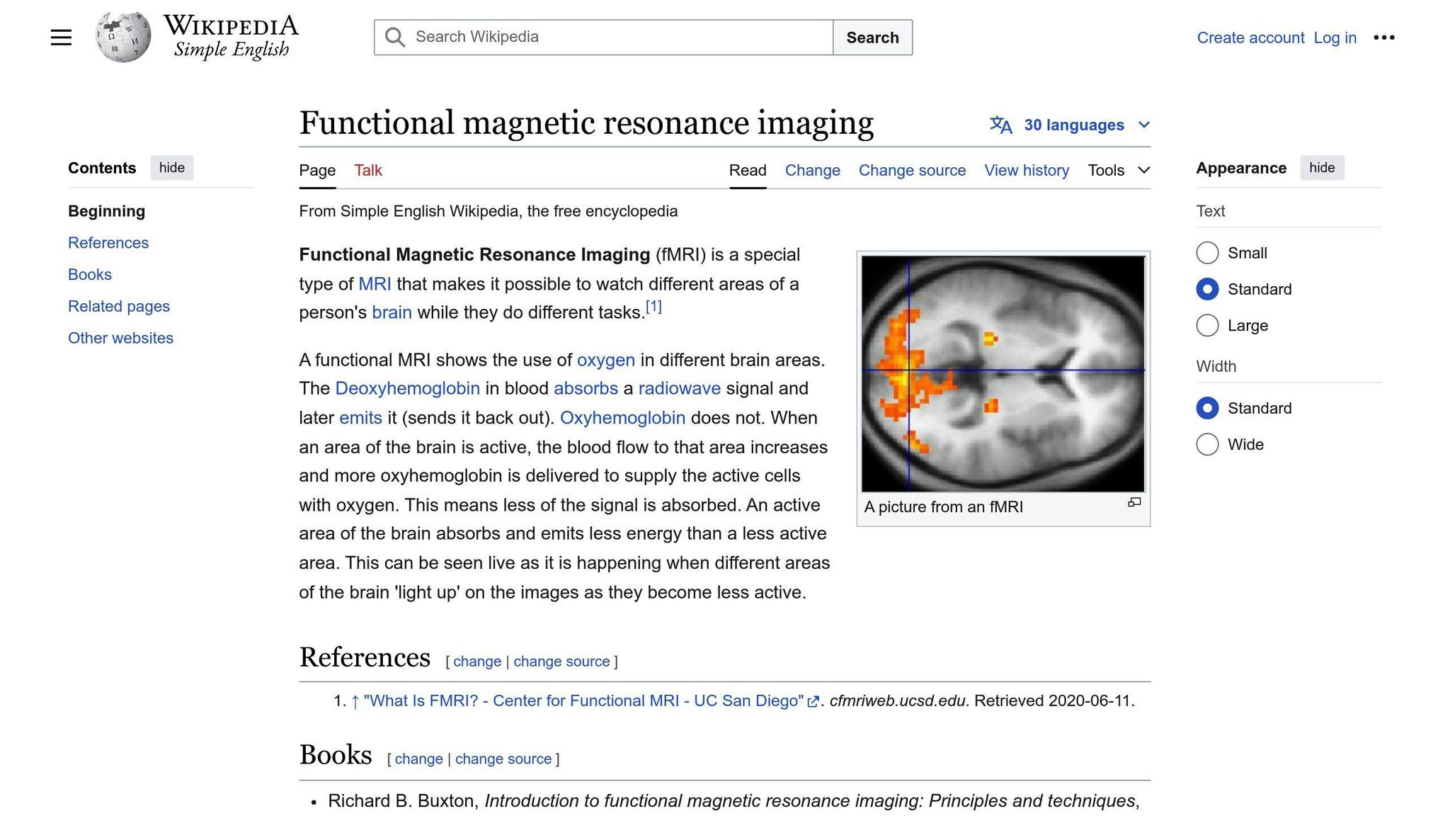
fMRI, or functional Magnetic Resonance Imaging, is a brain imaging method that shows brain activity in real-time. It works by detecting changes in blood flow and oxygen levels in the brain.
Here's how it works:
- When a brain area becomes active, it needs more oxygen.
- More blood flows to that area, bringing oxygen.
- fMRI detects this increased blood flow.
- The scanner creates images showing which brain parts are active.
fMRI uses strong magnetic fields and radio waves to create these images. It doesn't use radiation, making it safer than some other brain scans.
Pros and cons of fMRI
| Pros | Cons |
|---|---|
| Safe, non-invasive | Expensive |
| High spatial resolution (1mm detail) | Poor temporal resolution (5-second lag) |
| Shows brain structure and function | Requires complete stillness |
| More objective than questionnaires | Results can be hard to interpret |
fMRI vs. other brain scans
fMRI stands out from other brain imaging techniques:
- EEG (Electroencephalography): Measures electrical activity directly. Faster than fMRI but less precise in locating brain activity.
- PET (Positron Emission Tomography): Uses radioactive tracers. Good for measuring brain chemistry but involves radiation exposure.
- MRI: Shows brain structure. fMRI goes further by showing brain activity over time.
fMRI's ability to show both brain structure and function makes it a top choice for studying complex tasks like writing.
"Between 100,000 and 250,000 entries related to fMRI can be found in PubMed, indicating its extensive use in research."
This high number of studies shows how widely used and trusted fMRI has become in brain research.
Brain areas involved in writing
Writing is a complex task that engages multiple brain regions. Let's explore the main areas involved and the thinking steps in the writing process.
Main brain areas for writing
Several key brain regions work together during writing:
| Brain Area | Function in Writing |
|---|---|
| Left Premotor Cortex | Controls hand movements for writing |
| Superior Parietal Cortex | Spatial processing and letter formation |
| Broca's Area | Language production and grammar |
| Wernicke's Area | Language comprehension |
| Visual Cortex | Visualization of written content |
| Hippocampus | Memory retrieval for writing topics |
| Frontal Lobe | Planning and decision-making |
The left hemisphere plays a dominant role in writing for most people. A key region is Exner's area, located near the junction of the middle frontal and precentral gyri. This area is crucial for the memory of writing movements.
"The mastery of handwriting is based on the involvement of a network of brain structures specific to writing alphabet characters." - From an fMRI study on writing processes
Thinking steps in writing
Writing involves several cognitive processes:
- Idea generation: The frontal lobe and hippocampus work together to brainstorm and recall information.
- Language processing: Broca's and Wernicke's areas transform thoughts into words and sentences.
- Motor planning: The premotor cortex prepares hand movements for writing or typing.
- Execution: The motor cortex sends signals to muscles to perform the physical act of writing.
- Visual feedback: The visual cortex processes what's been written, allowing for corrections.
fMRI studies have shown that experienced writers use their brains differently than novices. For example:
"Good writers engage fewer neural regions to write a newly taught letter than poor writers, indicating a more efficient brain network."
This efficiency is particularly noticeable in the premotor and parietal cortices.
Interestingly, the method of writing also affects brain activity. Handwriting and typing engage different neural pathways:
"College students showed increased connectivity across the brain when handwriting words compared to typing them." - From a study at the Norwegian University of Science and Technology
This research suggests that handwriting may boost learning and memory more than typing, due to increased brain connectivity.
Key fMRI studies on writing
Main research findings
fMRI studies have shed light on the complex brain processes involved in writing. Here are some key findings:
1. Handwriting activates multiple brain regions
A study involving 12 young adults revealed that handwriting engages a network of brain areas, including:
- Left primary somatosensory and motor cortex
- Bilateral supplementary motor area (SMA)
- Pre-motor areas
The researchers found that different writing tasks activated these regions to varying degrees. For example, copying a sentence showed higher handwriting speed and less in-air time compared to copying a grocery list or phone numbers.
2. Self-generated writing boosts reading skills
Karin Harman James, Ph.D., conducted fMRI studies showing that self-generated actions, like printing letters by hand, are crucial for developing reading skills. This research highlights the importance of handwriting in educational settings.
3. Creative writing engages specific brain areas
An fMRI study with 28 participants examined the neural correlates of creative writing. The research found that:
- Brainstorming activated a parieto-frontal-temporal network
- Creative writing engaged motor and visual areas associated with handwriting
- The left inferior frontal gyrus (BA 45) and left temporal pole (BA 38) were linked to verbal creativity during writing
4. Typewriting vs. handwriting
A study comparing typewriting and handwriting in Japanese revealed:
- Both tasks activated common brain regions: left superior parietal lobule, left supramarginal gyrus, and left premotor cortex
- Typing showed greater activation in the left posteromedial intraparietal cortex
5. Writing experience affects letter perception
Research has shown that writing experience influences how the brain processes letters:
- The anterior left fusiform gyrus responds more to individual letters
- Adults who learned to form pseudoletters by hand showed increased activation in letter processing regions, unlike those who learned through typing or visual practice alone
How researchers study writing
fMRI researchers use various methods to study writing processes:
-
Task-based fMRI: Participants perform writing tasks during brain scanning. For example:
- Copying text (e.g., grocery lists, phone numbers, sentences)
- Creative writing tasks
- Brainstorming exercises
-
Comparative studies: Researchers compare brain activity during:
- Handwriting vs. typing
- Writing vs. drawing
- Writing vs. oral spelling
- Specialized equipment: Studies often use fMRI-compatible tablets or other devices that allow participants to write while in the scanner.
- Control tasks: Researchers include control tasks to isolate writing-specific brain activity.
- Individual differences: Some studies assess participants' writing skills or creativity levels to correlate with brain activity.
Brain activity during writing
Brain patterns in writing
fMRI studies have revealed complex brain activation patterns during writing tasks. Here's what researchers have found:
1. Multiple brain regions activate: Writing engages a network of brain areas, including:
- Left primary somatosensory and motor cortex
- Bilateral supplementary motor area (SMA)
- Pre-motor areas
- Left inferior frontal gyrus (BA 44/45)
- Left anterior insula
- Left temporal pole (BA 38)
2. Left hemisphere dominance: The left side of the brain shows stronger activation during writing tasks, especially in areas controlling the right hand.
3. Four key brain nodes: Studies consistently show activation in these regions during handwriting:
| Brain Region | Function |
|---|---|
| Dorsal premotor cortex | Motor planning |
| Ventral premotor cortex | Hand movements |
| Superior parietal cortex | Spatial processing |
| Fusiform gyrus | Visual word recognition |
4. Exner's area: This region, near the junction of the middle frontal and precentral gyri in the left hemisphere, is crucial for writing. It stores the memory of gesture sequences needed to produce each character.
What brain activity tells us
Brain activation patterns during writing tasks provide insights into the writing process:
1. Skill level differences: Good writers show more efficient brain activation, engaging fewer neural regions compared to poor writers. For example, in a study with children, good writers were more efficient when writing practiced letters, while poor writers showed no difference between new and practiced letters.
2. Writing method impacts: Different writing methods activate the brain differently:
- Handwriting activates almost the whole brain
- Typing engages fewer brain regions
A study by Professor Audrey van der Meer at NTNU found:
"Our main finding was that handwriting activates almost the whole brain as compared to typewriting, which hardly activates the brain as such."
3. Creative writing insights: An fMRI study with 28 participants showed:
- Brainstorming activated a parieto-frontal-temporal network
- Creative writing engaged motor and visual areas for handwriting
- The left inferior frontal gyrus (BA 45) and left temporal pole (BA 38) were linked to verbal creativity
4. Language processing: Writing activates brain areas associated with language, including Broca's Area and Wernicke's Area. These regions work together to process and produce written language.
5. Memory and visualization: When writing on physical paper, there's increased activity in areas linked to memory and visualization. A study by Professor Kuniyoshi L. Sakai at the University of Tokyo found:
"Our take-home message is to use paper notebooks for information we need to learn or memorize."
Understanding these brain activity patterns can help improve writing education and develop targeted interventions for individuals with writing difficulties.
sbb-itb-1831901
Different types of writing
Handwriting vs. typing
fMRI studies show that handwriting and typing engage different brain areas and processes. Here's what researchers have found:
- Brain activation: Handwriting activates almost the whole brain, while typing engages fewer brain regions.
- Memory and learning: Writing by hand enhances memory retention and learning compared to typing.
- Speed and efficiency: A study at the University of Tokyo found that participants who used paper completed note-taking tasks about 25% faster than those using digital devices.
- Brain connectivity: Handwriting stimulates more complex brain connections, which are essential for encoding new information and forming memories.
- Recall speed: People who wrote in paper calendars recalled information 25% faster than those who typed it into a smartphone.
| Aspect | Handwriting | Typing |
|---|---|---|
| Brain activation | Almost whole brain | Fewer regions |
| Memory retention | Higher | Lower |
| Task completion speed | Faster (25% quicker) | Slower |
| Brain connectivity | More complex | Less complex |
| Recall speed | Faster (25% quicker) | Slower |
Professor Kuniyoshi L. Sakai from the University of Tokyo states:
"Our take-home message is to use paper notebooks for information we need to learn or memorize."
Writing in different languages
fMRI studies have revealed how the brain processes writing in different languages:
- Native vs. second language: The brain handles native language (L1) and second language (L2) writing differently.
- Brain state dynamics: L1 processing is linked to more integrated brain states and greater transition flexibility. L2 processing involves more segregated states and less transition flexibility.
- Network integration: L1 processing uses a more globally integrated network, while L2 processing relies on a more segregated network.
- Comprehension: A study of Chinese-English bilinguals found higher comprehension scores for L1 (mean score = 37.82) compared to L2 (mean score = 32.50).
- Visual cortex adaptation: Research led by Laurent Cohen at the Paris Brain Institute examined how the visual cortex adapts to various writing systems, such as Latin and Chinese characters.
- Gender differences: A study on Chinese handwriting found that functional connectivity between Exner's area and the right cerebellum was greater in females compared to males.
These findings highlight the brain's flexibility in processing different writing systems and languages, which is crucial for bilingual individuals who master multiple writing methods.
Writing problems and fMRI
fMRI studies have shed light on how the brain functions during writing tasks, helping researchers better understand and diagnose writing disorders. These insights are crucial for developing targeted interventions for conditions like dysgraphia and dyslexia.
Common writing disorders
Two main writing disorders that have been studied using fMRI are dysgraphia and dyslexia:
| Disorder | Description | Brain Patterns |
|---|---|---|
| Dysgraphia | Impairs handwriting and spelling | Less white matter integrity in key brain areas |
| Dyslexia | Affects reading and spelling | Differential activation patterns during writing tasks |
A study led by Todd Richards at the University of Washington scanned the brains of 40 children diagnosed with dyslexia, dysgraphia, or who were typically developing readers and writers. The researchers found that:
- Typically developing children used fewer, more direct "highways" in their brain activity, indicating more efficient processing.
- Children with dyslexia and dysgraphia showed different patterns with fewer direct highways and more "detours."
These findings highlight the need for tailored instruction for specific learning disabilities. As Virginia Berninger, an educational psychologist involved in the study, stated:
"So many of these kids never get the kind of instruction they need. If you find out what's wrong and you teach that skill, voilà, they can learn."
Another study by Richards et al. (2015) compared brain activity in children with dysgraphia, dyslexia, and typical writers:
- The dysgraphia group showed less white matter integrity in key brain areas compared to typical writers.
- Poor writers activated more brain regions than good writers during writing tasks, suggesting less efficient neural processing.
This research has practical implications for diagnosis and treatment:
- Early identification: fMRI can help identify writing disorders earlier, allowing for timely intervention.
- Targeted interventions: Understanding the specific brain areas affected can lead to more focused treatment approaches.
- Progress monitoring: Brain scans can potentially track improvements in neural efficiency as children undergo writing interventions.
It's worth noting that writing disorders are not uncommon. About 36 percent of students receiving special education services in the United States have normal intelligence but struggle with reading, writing, speaking, and doing math.
New methods and future research
Recent advancements in fMRI technology are opening new doors for studying writing processes in the brain. These innovations allow researchers to capture brain activity with greater precision and speed, paving the way for more detailed insights into the neural mechanisms of writing.
New fMRI techniques
1. Ultra-fast brain activity measurement
Researchers have developed a technique that measures brain activity 60 times faster than traditional fMRI. This method can track neural responses within 100 milliseconds of stimuli, with early data suggesting it may detect changes as fast as 24 milliseconds.
"The intriguing novelty of this approach is that the stiffening and softening of specific brain regions persists even when stimuli as short as 100 milliseconds are presented to the mouse." - Sam Patz, Physicist, Brigham's Department of Radiology
This technique, based on magnetic resonance elastography (MRE), creates maps of tissue stiffness using an MRI scanner. It has shown that the auditory cortex in anesthetized mice stiffens in response to stimuli, indicating a link between brain stiffness and neuronal activity.
The NexGen 7T scanner offers ultra-high resolution imaging, allowing researchers to study brain circuitry with greater detail. Key features include:
| Feature | Capability |
|---|---|
| Resolution | Up to 10 times more detail than current 7T scanners |
| Voxel size | Can see fMRI features as small as 0.4 millimeters |
| Neuron detection | Can detect activity in about 850 individual neurons within a single voxel |
This technology enables the study of brain activity at different depths in the cortex, helping to reveal intricate brain circuitry by differentiating activity in various cell layers.
3. SPICE technology
Fan Lam's research team has developed SPICE (spectroscopic imaging by exploiting spatiospectral correlation), an advanced MR spectroscopic imaging (MRSI) technique. This technology allows for:
- Whole brain mapping of metabolites in just five minutes
- Resolution matching that of a standard functional MRI scan
- More than tenfold improvement over existing methods
SPICE adds another dimension to standard MRI exams by mapping biochemical profiles of the brain at high resolution.
Future research ideas
1. Exploring writing disorders
Future studies could use these advanced fMRI techniques to better understand writing disorders like dysgraphia and dyslexia. The increased spatial and temporal resolution might reveal subtle differences in brain activity patterns between typical writers and those with writing difficulties.
2. Investigating the impact of different writing methods
Researchers could use ultra-high resolution fMRI to compare brain activity during handwriting versus typing, potentially uncovering new insights into how different writing methods engage various brain regions.
3. Studying the writing process in real-time
The ultra-fast fMRI techniques could allow researchers to observe brain activity changes as a person progresses through different stages of the writing process, from ideation to execution.
4. Cross-linguistic studies
Advanced fMRI methods could be used to compare brain activity patterns in individuals writing in different languages or scripts, potentially revealing how the brain adapts to various writing systems.
5. Machine learning integration
Incorporating machine learning techniques with high-resolution fMRI data could uncover hidden patterns in neural activity during writing tasks, potentially leading to new diagnostic tools or therapeutic approaches for writing disorders.
As these new fMRI methods continue to evolve, they promise to deepen our understanding of the complex neural processes involved in writing, potentially leading to improved diagnostic and therapeutic strategies for writing-related challenges.
Conclusion
fMRI studies have provided valuable insights into the complex neural processes involved in writing. These studies have revealed key differences between good and poor writers, shedding light on the brain's role in various aspects of written expression.
Key findings from fMRI research on writing include:
- Brain activation patterns: Good writers show more efficient brain activation compared to poor writers. Poor writers tend to use more brain areas to complete the same tasks, indicating less efficient neural processing.
- Specific brain regions: The left fusiform gyrus plays a crucial role in orthographic coding (handwriting). Activity in the left posterior cingulate, left precuneus, and right precuneus regions correlates with spelling and written composition skills.
- Task complexity: Brain activity increases with the complexity of writing tasks. For example, copying sentences engages more language processing areas compared to simpler tasks like copying phone numbers.
- Learning new letter forms: Poor writers show more extensive brain activation when learning new letter forms, suggesting inefficiency in this process.
- Handwriting impairments: fMRI has been used to study handwriting impairments in conditions like Alzheimer's disease, providing a foundation for future research on brain dysfunctions affecting writing.
These findings have practical implications for education and therapy:
| Application | Potential Benefit |
|---|---|
| Educational interventions | Targeted strategies to improve writing skills based on brain activation patterns |
| Diagnostic tools | Early identification of writing difficulties using fMRI-based assessments |
| Therapeutic approaches | Development of brain-based therapies for writing disorders |
As fMRI technology advances, researchers are gaining more detailed insights into the writing process. New techniques, such as ultra-fast brain activity measurement and high-resolution imaging, promise to further our understanding of the neural mechanisms underlying writing.
Future research directions may include:
- Exploring the neurobiological basis of specific writing disorders
- Investigating the impact of different writing methods (e.g., handwriting vs. typing) on brain activity
- Studying cross-linguistic differences in writing processes
FAQs
What parts of the brain are used for writing?
Writing isn't just about putting pen to paper. It's a complex process that engages multiple brain regions. Here's a breakdown of the key areas involved:
| Brain Region | Function in Writing |
|---|---|
| Left premotor cortex | Controls the motor aspects of writing |
| Left superior parietal cortex | Manages spatial aspects and hand movements |
| Left fusiform gyrus | Processes visual word forms |
| Angular gyrus | Integrates information for handwriting and reading |
But that's not all. A recent meta-analysis identified 12 distinct areas that light up during writing tasks. The standout regions? The posterior part of the left superior frontal sulcus (aka the graphemic/motor frontal area or GMFA) and the left superior parietal cortex (SPL).
These areas aren't just active; they're writing superstars. They show a stronger response to writing compared to other motor or language tasks. It's like they have a special love for penmanship.
Here's the kicker: If these areas get damaged, it can lead to some serious writing issues. For example, lesions in the left frontal superior or parietal superior regions can cause pure agraphia syndrome - a condition where a person can't write but can still read and speak just fine.